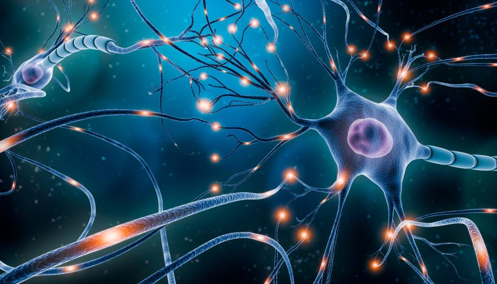
Given the recent clinical interest in psychedelic compounds, many researchers have sought to elucidate their mechanism of action from a neurophysiological perspective. These research teams are seeking to understand changes in brain activity between the psychedelic state and normal waking consciousness. More specifically, the goal of these studies is to characterize the networks and regions of the brain affected by psychedelic ingestion. This can be accomplished with functional magnetic resonance imaging (fMRI), which accurately produces real-time depictions of brain activity.1
Effects of Psilocybin on the Default Mode Network
A seminal 2012 study at Imperial College London was the first to use fMRI to characterize brain activity between psychedelic and non-psychedelic states.2 In this work, Carhart-Harris et al. used fMRI to record alterations in brain activity before and after injections of placebo and psilocybin. The paramount discovery was that psilocybin appeared to reduce overall cerebral blood flow. The brain regions which demonstrated the most consistent deactivation were the posterior cingulate cortex and the medial prefrontal cortex. These two regions reside in the default mode network (DMN), which refers to specific areas of the brain involved in various domains of cognitive and social processing, as well as autobiographical memory recollection.3,4
In normal waking consciousness, activity in the posterior cingulate cortex and medial prefrontal cortex is relatively high compared with other regions of the brain. Interestingly, the degree to which activity in these regions was decreased was proportional to the intensity of the subjective effects reported by the volunteers. There is speculation that the posterior cingulate cortex is involved in consciousness, formulation of the self, and ego.5-8 The “ego-dissolution” phenomenon is a commonly reported experience following the administration of high doses of psychedelics. While this could be related to decreased posterior cingulate cortex activity, no direct quantitative relationship has been observed between the disintegration of the DMN and ego-dissolution.
A study published earlier this year from the University of Zurich also examined psilocybin-induced neurophysiological changes in volunteers.9 Like the previously mentioned study, this group used fMRI to capture changes in brain activity as the psilocybin-induced psychedelic state began to manifest in their volunteers. Their results supported the findings of the group at Imperial College London, in that associative regions become disintegrated with the rest of the brain upon psilocybin ingestion. Additionally, this study found that sensory areas of the brain, which are related to motor function, sensation, and visual perception become more integrated with the rest of the brain in the psychedelic state.
LSD Demonstrates Similar Effects on the Brain
The team at the University of Zurich also conducted a similar study with LSD, another tryptamine-derived hallucinogen.10 Observations regarding the neurophysiological effects of LSD are consistent with the two previously mentioned studies using psilocybin. LSD ingestion results in a decreased connectivity of associative regions of the brain, and increased integration of sensory and somatomotor regions. Moreover, participants who showed relatively higher connectivity in the somatomotor network also reported a higher intensity of subjective effects.
How Do Tryptamine Hallucinogens Affect the Whole Brain?
A study from the Institute for Scientific Interchange in Turin, Italy indicates that psilocybin induces more persistent connections throughout the brain as a whole compared with normal waking consciousness.11 This work was conducted using the original fMRI data collected at Imperial College London. The researchers say psilocybin induces a more intercommunicative mode of brain function. This may mean there is more sharing of information between brain regions that don’t associate with each other in normal waking consciousness. Furthermore, the researchers speculate that this intercommunicative mode of brain function may be the root of the synesthesia phenomenon associated with the psychedelic experience.
Summary
Collectively, these studies help describe the neurobiological mechanisms of tryptamine-derived hallucinogens. The general observation is higher-level functional networks involved in various cognitives processes become less integrated, and sensory and somatomotor networks become more integrated. What’s more, there seem to be more persistent connections formed throughout the whole brain during the psychedelic state relative to normal waking consciousness. These studies add to the psychedelic knowledge base which other researchers can tap into for further investigation.
More persistent brain connections? How fascinating!
Are more persistent connections related to “Hallucinogen Persisting Perceptual Disorder” (HPPD), and is HPPD associated with not allowing tolerance to subside with serial experiences, or worse, overcoming tolerance with increased dosage? All of this leading to increased time in the psychedelic state, facilitating increased connections to become “normalized?”